The COVID-19 pandemic has created long-term disruptions for drug development programs. Clinical research visits present exposure risks to patients with high-risk conditions. Healthcare systems have redirected resources to essential support for COVID-19 treatment and research. As a result, many clinical trials outside of COVID-19 research have slowed, stalled, or stopped. Ongoing studies have encountered major challenges in the already difficult clinical trial recruitment process as patients have deferred healthcare visits and are hard to identify as potentially qualified participants or unavailable for engagement.
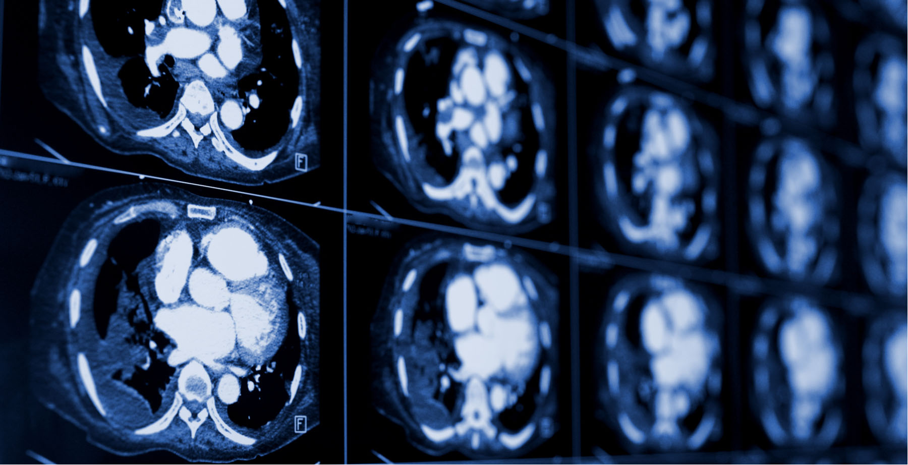
Just as healthcare providers have moved to telehealth, life sciences organizations are seeking to transition to virtual or contactless methods to progress critical path research if not permanently shift research into a future-state of remote monitoring. Real-world imaging, or the incorporation of diagnostic images into real-world evidence (RWE) programs, provides multiple solutions to accelerate clinical research including new ways to construct regulatory submissions, algorithms to identify patients for recruitment, and modifications to protocol design.
Imaging is Key for New Observational Regulatory Submissions
Achieving approvals from the Food and Drug Administration (FDA) for Investigational New Drugs (IND) or marketing label indication expansion on approved products require detailed data sets sufficient to support regulatory decisions on safety and efficacy. Section 3022 of the 21st Century Cures Act, enacted in December 2016, directed the FDA to evaluate RWE as a tool to support approvals for new drug indications. The following year, “A Framework for Regulatory Use of Real-World Evidence” published by Duke-Margolis Center for Health Policy opined that observational studies may be capable of “generating evidence for a label change that incorporates new information closely associated with the accepted clinical evidence in the label or makes revisions to that information.” These efforts opened the door for label expansion to new indications through real-world data (RWD) without an interventional study and to employ synthetic controls from RWD as a component of regulatory submissions of interventional studies.
Multiple life sciences companies have since employed observational approaches in their submissions. An early example is Amgen’s Blincyto for leukemia that was approved in 2018 through data submitted from observational studies. A recent study by Genentech detailed applying synthetic controls using electronic health records to derive control arms for single‐arm lung cancer trials. Research continues to establish that synthetic controls can generate sufficient data and may reduce the need for control placebo patients in randomized controlled trials (RCT) from 1:1 to 3:1 or 5:1.
Maturing RWE Programs are Vital
Maturing capabilities in RWD programs are vital given the lack of direct patient engagement and in-person visits that resulted from COVID-19. Interventional randomized clinical trials collect a high standard of information from each patient case using a combination of standardized study visits, detailed annotation of imaging and pathology studies, and case report forms that standardize data into controlled vocabularies and comparable values.
Real-world imaging has become a critical component of these programs because radiology is among the core areas where the management of data is different between the level of annotation captured in care delivery and clinical research. For example, the Response Evaluation Criteria in Solid Tumors (RECIST) methodology used to annotate cancer cases provides regulatory research-grade information to track response to therapy as measured in PET imaging studies. While solid tumor response is tracked in routine cancer care, the RECIST criteria are not regularly applied to clinical trial cases. In order to support research for a regulatory submission, RECIST annotations need to be abstracted or automatically determined from historical cases. Having the original images from each patient follow-up visit is essential to making this work.
Oncology isn’t the only area where radiology is one of the most important mechanisms to determine disease progression. Cardiology and pulmonology teams routinely use CT imaging to determine properties of the heart, such as ejection fraction, and details in various tissues in response to therapy. Neurology researchers viewing conditions such as Multiple Sclerosis or Parkinson’s disease review lesions and neuroanatomical changes. Even when the existing free text radiology reports are sufficient for providing findings of interest, a regulatory-grade submission differs from a comparative effectiveness or health economics study.
Traditional comparative effectiveness research (CER) and health economics outcomes research (HeOR) studies are used in reimbursement decision-making or to guide prescribing behavior but do not directly control the FDA label. For IND applications and label expansions, the data must be submitted to the FDA and data must meet GxP compliance standards to ensure products are safe, meet their intended use, and adhere to quality processes. This translates to requirements for a record that can be audited and traced to the raw source data that needs to be made available on request or included in the submission. Having access to the original imaging data files and audits to validate accuracy of interpretations within radiology studies used in the submission are still expected for FDA submissions, making it important to have a real-world imaging resource that can meet these requirements.
Machine Learning to Support Trial Planning and Patient Recruitment
A major achievement in the past decade is the advancement of reliable and scalable machine learning to automatically classify objects and features in images. Clinical trial recruitment involves identifying patients with the right characteristics for both inclusion and exclusion criteria. While patients may not be visiting doctors as frequently as in the past, their records include a large volume of clinical data that can be used for improving the clinical trials. As clinical development teams review trial designs to adapt to COVID-19 shifts, they can look to historical records to model factors that could be identified by data and logic rather relying on clinical teams to scout patients.
RWD is essential for understanding how to enhance the next generation of studies including how to optimize for measurement in a new remote paradigm. Data that can be established through remote monitoring or biomarkers, and can provide quantitative end points that can be captured within imaging studies, can move trials forwards with a lower reliance on clinical visits or establish evidence with smaller experimental sizes or compressed study timelines. Real-world imaging provides the basis for defining adjustments to study cohort definitions, data collection requirements, sensitivity of research tools, and biomarkers for end points.
AI tools that allow consumer applications to recognize faces are highly applicable to analyzing radiology images. Historical radiology studies and pathology studies in conjunction with medical records can be used to train algorithms to identify patients who meet clinical trial criteria. This mechanism can be especially useful for rare diseases that are inconsistently diagnosed because the symptoms appear similar to clinical teams. Even standard radiology studies can show very specific anatomical or biological abnormalities that indicate a potential case. Life sciences teams interested in the potential of radiology are investing in acquiring data sets to train novel algorithms and will share those algorithms with teams at clinical sites to support patient identification or exclusion as recruitment ramps up again. For undiagnosed patients, some teams are looking at the benefits of using historical data on patients to expand the market for products by improving the diagnostic process with machine learning algorithms trained on real-world imaging data sets. These groups are working towards building the algorithms today and will work towards deploying them to engage patients directly in the coming years.
Although the clinical development process has been disrupted by COVID-19, this pause is an opportunity to innovate in areas such as study efficiency and expanding markets. Radiology data sets are increasingly available with growing volume from data processors and are being integrated into study-grade content through digital contract research organizations (CRO). Now is the time to invest in real-world imaging to pilot or scale drug development programs.


