When medical device companies want to demonstrate how their device works, they pull out a potato or a bell pepper and act if that is the organ on which the device will be used. The only problem with that—vegetables aren’t organs. Fortunately, Lazarus 3D, a new startup, can 3D print lifelike organs that can be used for medical education, including testing and demonstrating medical devices as well as helping surgeons practice before actual operations.
Founder and President Jacques Zaneveld, PhD, developed this revolutionary new technology, which allows the company to 3D print using extremely soft material. Even better, they can create copies of patient body parts from real medical data in order to provide custom models of real patients for practice prior to actual surgeries. PM360 spoke with Dr. Zaneveld about how the company got its start, working with medical device companies, and what’s next for Lazarus 3D.

PM360: Where did the idea come from to start this company?
Dr. Jacques Zaneveld: It started when I was a PhD student at Baylor College of Medicine where I built my first 3D printer. Originally, I went to anime conventions every weekend to find some way to sell 3D-printed stuff, such as little figurines and throwing stars.
But then I participated in a study on how the brain works with some of my fellows at Baylor College of Medicine, in which they scanned my brain. From that scan, I built a model of my own brain, which I showed to a few people, including a medical expert for lawsuits involving personal injury. After that, I started making models of people who reported they were injured as part of lawsuits to help demonstrate the injury to the jury.
But what I really wanted to do was find a way to apply it to medicine. So, I talked to a few of the doctors at the medical school and one of them, Dr. Richard Link, said we should figure out a way to make lifelike copies of body parts so that they could rehearse upcoming operations.
How realistic are these models?
Once I was able to reliably and accurately reproduce lifelike replicas of body parts, I was able to get a sample into a surgical setting within a couple days—and the doctors loved it. They said it was more realistic than any of the models that were available on the market. Later, I published a study with Dr. Link’s group in December 2016 in which we describe 13 different cases that showed real operations were more effective when you made a model of that patient and then rehearsed on it. In fact, in one of those cases, it seems very likely that the patient was able to retain their kidney because the doctors were able to practice.
How did you get the funding to start the company?
I started out with $10,000 that a fellow post doc at Baylor College of Medicine gave me. That helped pay for the first few months of rent as well as salary for the first employee that I hired. And basically, from day one we’ve had sales. We’re very lucky in the fact that we partnered with a lot of forward-looking medical institutes and medical device companies that have been willing to pay for our services.
What are medical device companies using these 3D replicas for?
A lot of medical device companies have a couple of very pressing problems. First, they have to convince doctors that the devices they produced work and are going to be successful in a real operation. Second, they need to train doctors so that they know how to properly use those devices to maximize success rates.
For example, when I went to the American Association of Gynecologic Laparoscopists meetings in D.C., major billion-dollar companies were using things such as bell peppers, pumpkins, and potatoes to show how their medical devices work. And quite frankly, that’s pathetic—we can do much better than that.
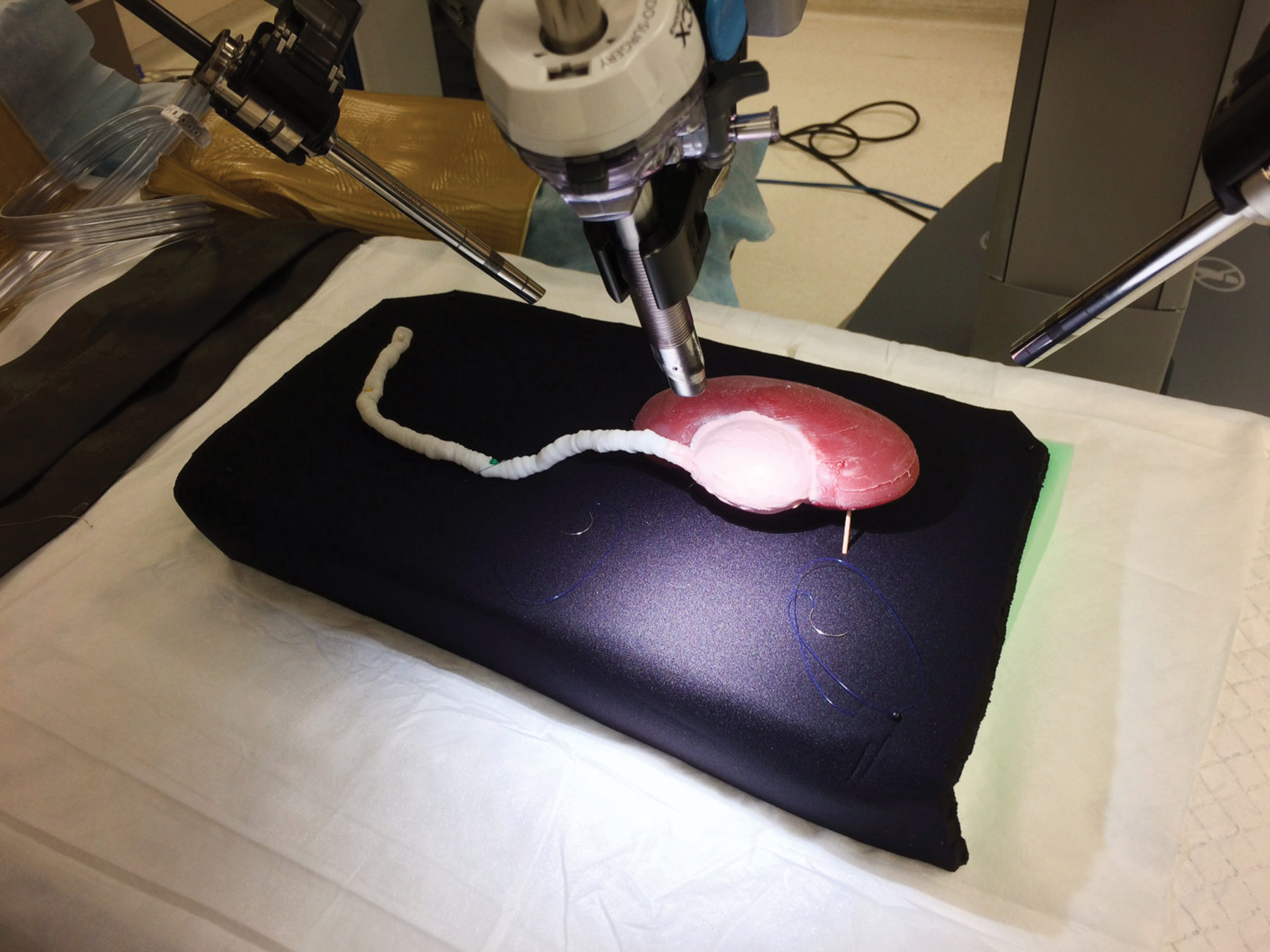
Since we can customize our models, we can make a model that you can use to show in a very realistic way exactly how your device works. You can demonstrate it right in front of doctors without needing to schedule expensive and complicated cadaver labs or animal labs. A lot of companies have found that it is a very powerful tool for marketing.
We also work with the research and development arms of various medical device companies. So, we’ve saved a lot of pigs’ lives, because rather than sacrificing a pig every week to try out a new iteration or component of a device, companies can instead have one of our benchtop models. Not only is this more efficient and effective, but data from these models can be submitted to the FDA for a consideration as your development process moves forward.
Can you produce a model for any organ?
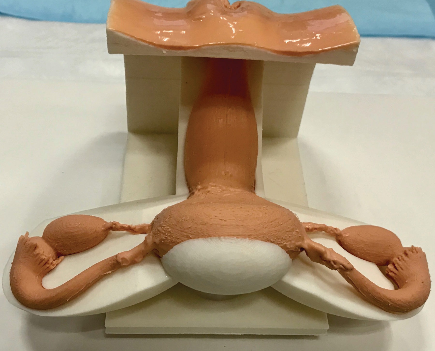
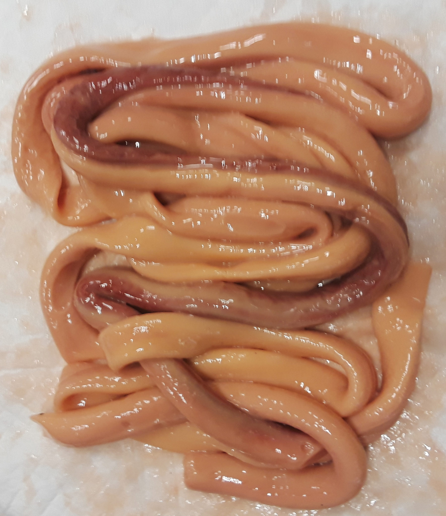
Yes, absolutely. Just within the past couple of weeks we’ve made models of the intestines, liver, kidneys, brain, skull, bladder, and reproductive systems. We recently got an order for a jaw, to which we can add very realistic gums and soft tissue. We can also do internal architecture, such as arteries, veins, the biliary system, or whatever might be required.
What are your future plans for the company? Do you ever intend to make live, workable organs?
It will take some time and significant amounts of investment before we get to a point where we can make organs that are regularly transplanted into human beings. So, as an early-stage company, my only focus is on making models that can accurately be used for medical education and practice in surgery.
Right now, we are considering our first large capital raise. And we are very excited to meet anyone who would like to partner up with us on this adventure. I believe that there is a multi-billion-dollar market in patient-specific modeling. For every 1% of surgery where a model could be used, you’re looking at about $500 million in potential revenue.
Examples of Lifelike 3D Printed Organs from Lazarus 3D
Female Reproductive System
Intestines
Cesarean Section
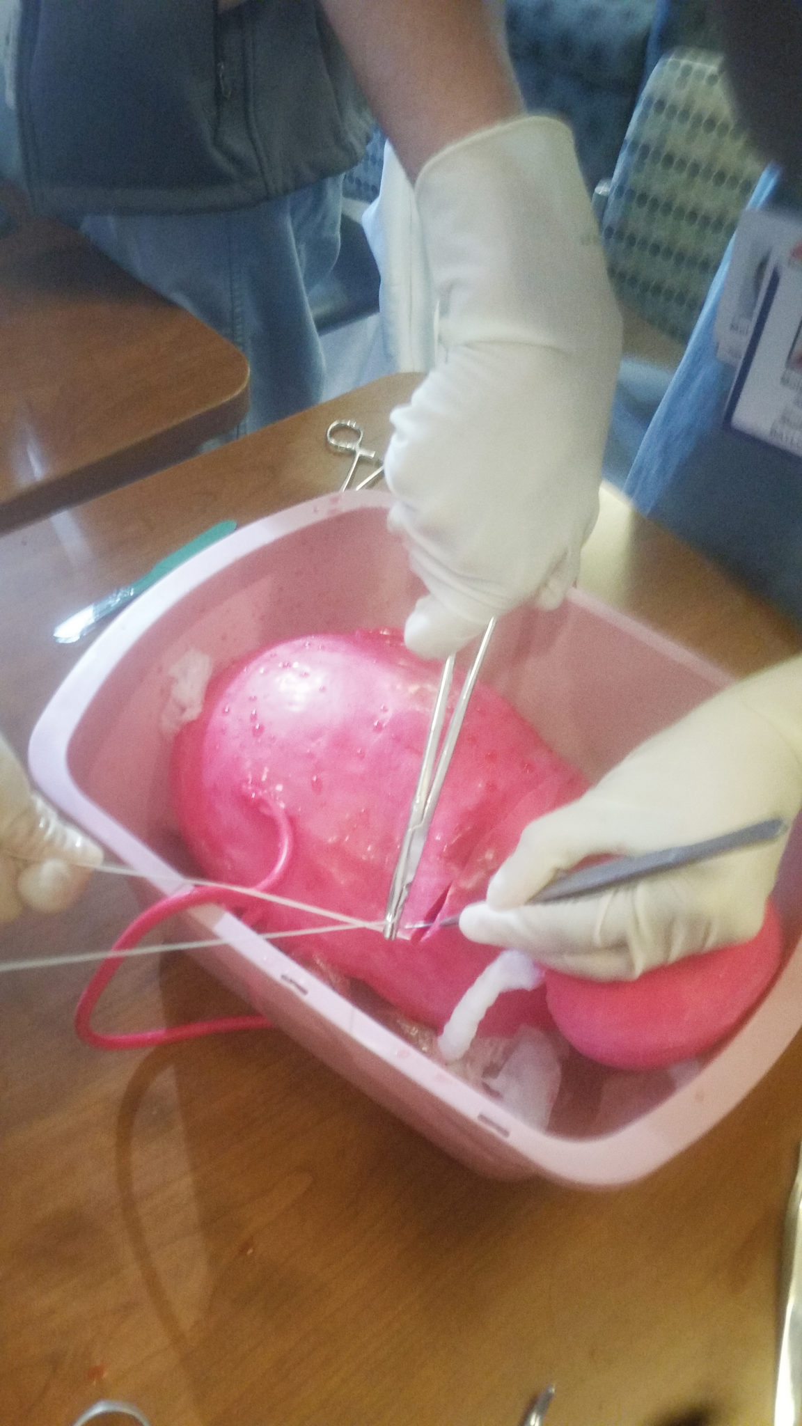
Kidney and Ureter